Increased precision in surgery thanks to our innovative simulation and design process.
We provide bespoke solutions for surgeons who put their patients at the core of their care by using the latest 3D technologies to create bespoke Cranio-maxillofacial implants, sub-periosteal implants, Temporomandibular joint, custom-made artificial joints, extra aural devices, intra and extra devices, artificial heart valves and spinal implants.
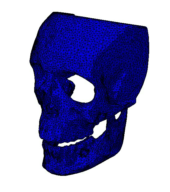
These 3D models are a great asset for surgeons as it allows them to visualise the region before an upcoming surgery, meaning they have more information before entering the operating theatre as the models are used to simulate surgery due to the possibility of being able to drill and cut into the models.
How our 3D models benefit you...
Plan the Procedure more accurately
Being able to view the patient's exact anatomy allows surgeons to understand their requirements in surgery better.
Surgical Simulation
The ability to carry out surgical simulations on the models means allows the official procedure to run smoothly, faster and more precisely.
High Resolution
Allows surgeons to view the exact structure of the patient, including the ability to distinguish teeth, grafts and nerves.
Improves Patient and Doctor Communication
Helps communicate plainly with patients and their family who don't understand the technical language, so is useful for demonstrating the procedure.

How do we create patient specific 3D anatomical models?
At Attenborough Medical, our service works by receiving the individual patient's CT or CBCT scan of the region of interest. We'll then 3D print your model(s) using our highest accuracy 3D printer. Our models are suitable for laboratory or for use in the operating theatre when not used by the scrubbed team.